Laboratory technology · reviewed
Electron microscopy
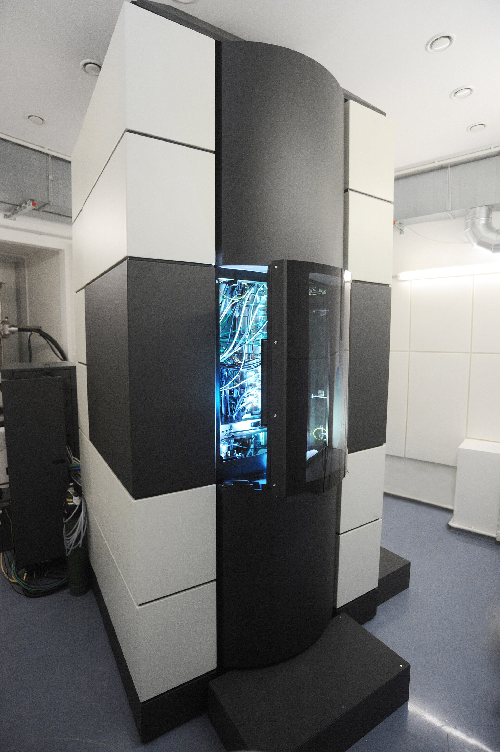
From 1931Electron microscopes use electron beams and electromagnetic lenses to resolve structures below the light-microscope limit.
Knoll and Ruska built early instruments in 1931–32. Biological applications expanded in the 1940s and 1950s as preparation and contrast methods revealed cellular ultrastructure and viruses. Because electrons have much shorter wavelengths than visible light, these instruments can reveal membranes, organelles, viruses, and other extremely small structures. The trade-off is demanding specimen preparation: samples are generally examined in a vacuum and are not viewed as living tissue.
Sources
- Tools of Cell Biology — Geoffrey M. Cooper, 2000
- Diagnostic electron microscopy is still a timely and rewarding method — Hans R. Gelderblom and Joachim Reupke, 1999
Direct relationships
- Vacuum tube→Electron microscopySupplied vacuum and electron-beam engineering precedents · high confidence
- Microscopy→Electron microscopyExtended microscopic imaging beyond visible-light resolution · medium confidence
- Electron microscopy→Molecular diagnosticsProvided complementary structural identification in specialized diagnostics · low confidence