Guided story · medical imaging
Seeing inside
Follow three connected routes—from electrical experiments and X-rays to ultrasound, computed tomography, and magnetic resonance imaging.
Radiation becomes a diagnostic tool
Electrical science and vacuum apparatus created the conditions for a new kind of penetrating radiation—and clinicians rapidly found a use for it.
- From 1820
Electromagnetism
Electric currents and magnetic fields were shown to be physically linked in the nineteenth century.
Locate in graph → - From the 1890s; electronic valve patented 1904
Vacuum tube
Vacuum tubes made it possible to direct, rectify, and amplify electron flow.
Locate in graph → - 1845–1923
Wilhelm Röntgen
Röntgen reported a new penetrating radiation in 1895 and demonstrated its imaging potential.
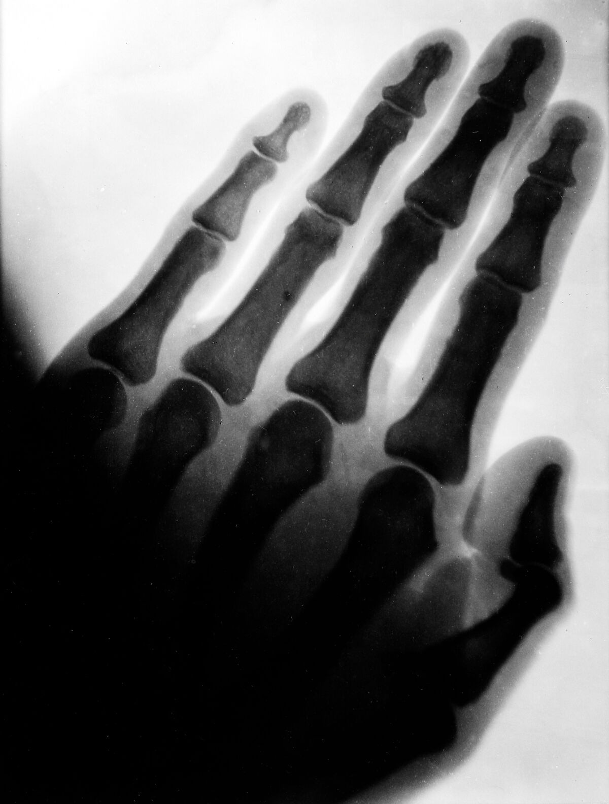
Locate in graph → - 1895
X-rays
X-rays made internal structures visible by recording differences in tissue attenuation.
Locate in graph → - From 1896
Early radiography
Clinical radiography followed within months of Röntgen's announcement.
Locate in graph → - 1867–1934
Marie Curie
Curie organized mobile radiography and trained operators during the First World War.
Locate in graph →
Echoes reveal living structures
A different branch of physics converted pressure into electrical signals, carrying pulse-and-echo methods from engineering into real-time clinical imaging.
- 1880
Piezoelectricity
Some crystals convert mechanical stress into electric charge and electrical excitation into motion.
Locate in graph → - From the 1910s
Sonar
Echo-ranging systems developed for navigation and warfare established techniques later adapted to medicine.
Locate in graph → - From the 1940s
Medical ultrasound
Ultrasound forms images from echoes of high-frequency sound transmitted into the body.
Locate in graph →
Electronics turn measurements into images
Solid-state electronics and computation allowed instruments to reconstruct slices, process weak signals, and distinguish tissues in new ways.
- From 1947
Semiconductor transistor
The transistor provided compact, low-power amplification and switching without heated vacuum tubes.
Locate in graph → - From 1971
Computed tomography
CT reconstructs cross-sectional images from many X-ray measurements around the body.
Locate in graph → - From 1973
Magnetic resonance imaging
MRI uses magnetic fields, radiofrequency signals, gradients, and computation to map tissue properties.
Locate in graph →