Scientific discovery · reviewed
X-rays
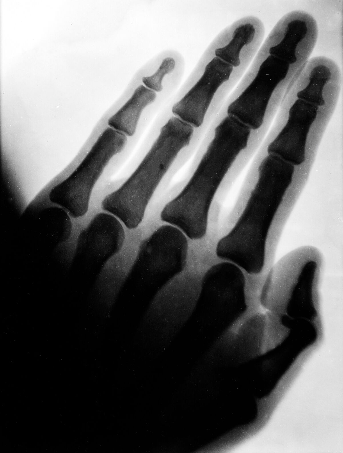
1895X-rays made internal structures visible by recording differences in tissue attenuation.
Discovered in 1895, X-rays were quickly used to image fractures and foreign objects. Their diagnostic value is substantial, but ionizing radiation also requires dose control and protection. Dense materials such as bone absorb more of the radiation than surrounding soft tissue, producing contrast on a detector. This made diagnosis possible without surgery, but the same ionizing energy can damage cells, so modern imaging balances useful information against radiation exposure.
Sources
- Medical Imaging: From Roentgen to the Digital Revolution, and Beyond — Hedvig Hricak et al., 2018
Direct relationships
- Vacuum tube→X-raysDischarge tubes provided the apparatus in which X-rays were observed · medium confidence
- Wilhelm Röntgen→X-raysReported and characterized X-rays in 1895 · high confidence
- X-rays→Early radiographyTurned penetrating radiation into diagnostic photographs · high confidence
- X-rays→Computed tomographyProvided CT's measured transmission signal · high confidence