Clinical technology · reviewed
Medical ultrasound
From the 1940sUltrasound forms images from echoes of high-frequency sound transmitted into the body.
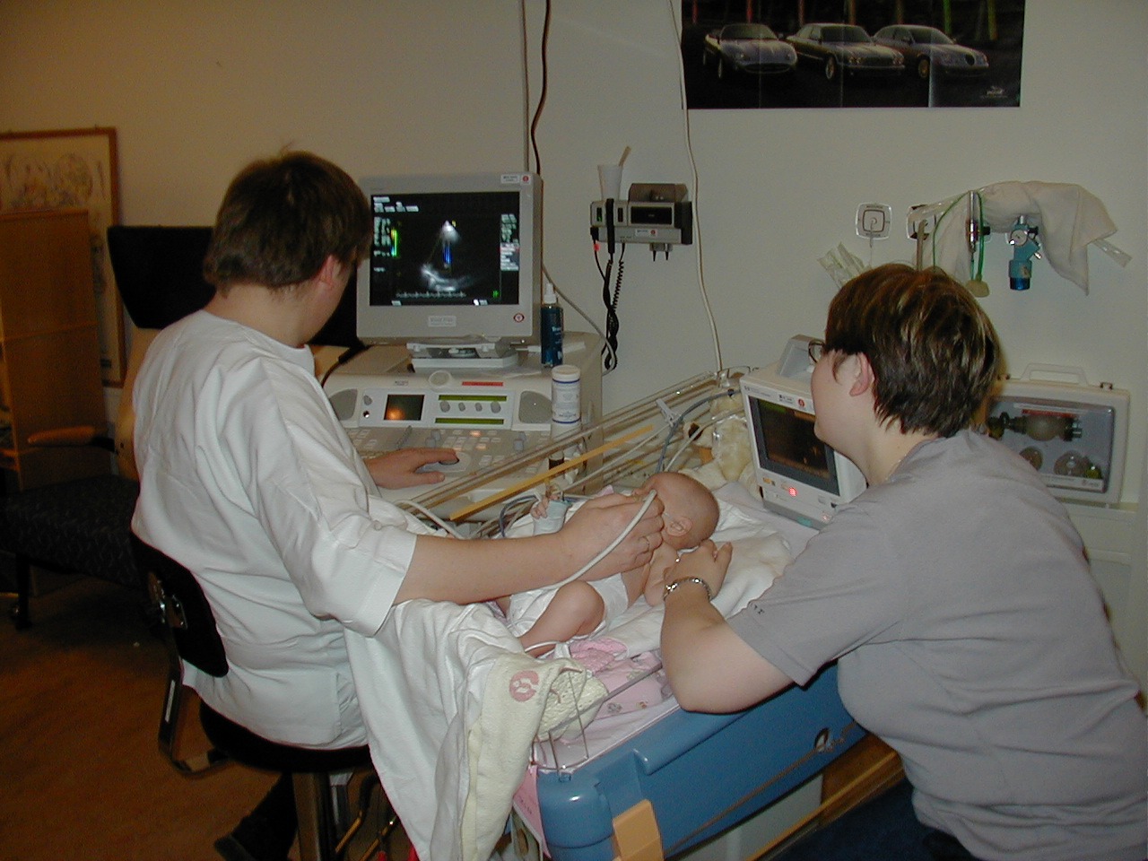
Clinical experiments began in several countries during the 1940s and 1950s. Piezoelectric transducers, pulse-echo methods, amplification, and display electronics gradually made real-time, non-ionizing imaging practical. A probe sends short sound pulses into the body and listens for echoes from tissue boundaries. Because it produces moving images without ionizing radiation, ultrasound became especially useful in obstetrics, cardiology, abdominal imaging, and procedures that require real-time guidance.
Sources
- My ultrasound used to look like what? — Lauren Rever, 2017-03-10
- Medical Imaging: From Roentgen to the Digital Revolution, and Beyond — Hedvig Hricak et al., 2018
Direct relationships
- Piezoelectricity→Medical ultrasoundEnabled ultrasound transmission and echo reception · high confidence
- Sonar→Medical ultrasoundProvided pulse-echo engineering precedents · medium confidence
- Semiconductor transistor→Medical ultrasoundMade amplification, processing, and compact display systems practical · medium confidence