Laboratory technology · reviewed
Microscopy
From the 17th centuryMicroscopes opened cells and microorganisms to systematic observation.
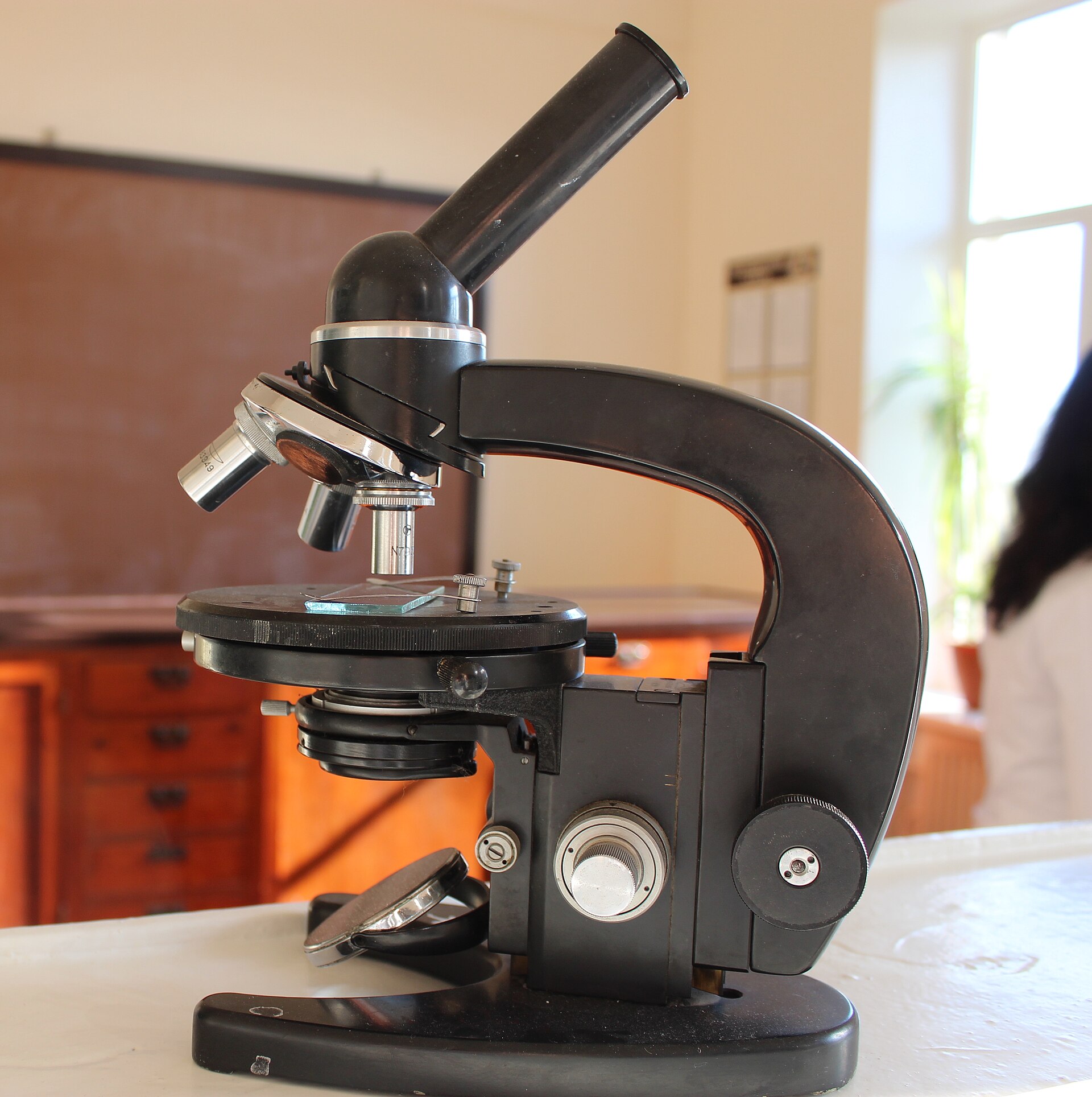
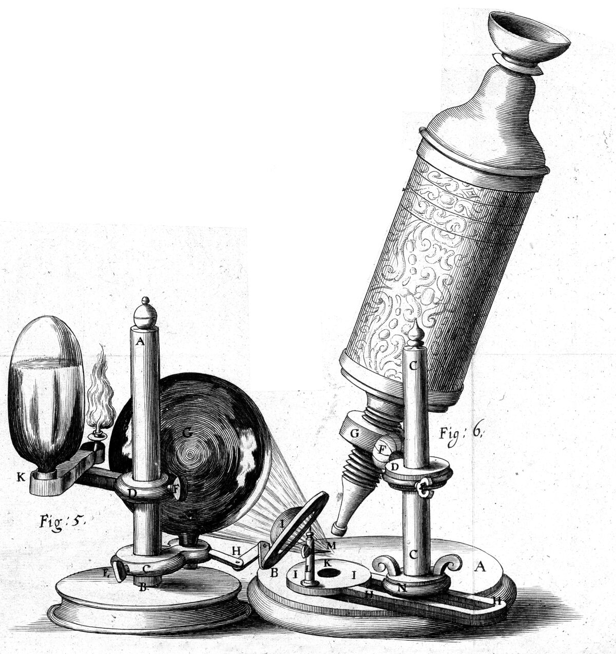
Hooke described cork compartments as cells in 1665, and Leeuwenhoek observed living cells and bacteria in the 1670s. Continued optical and specimen-preparation advances made microscopy fundamental to biology and pathology. Magnification alone was not enough: improved lenses, illumination, staining, and specimen preparation gradually made microscopic observations more reliable. Microscopy transformed medicine by connecting symptoms and visible tissue changes to cells, microorganisms, and the fine structure of organs.
Sources
- Tools of Cell Biology — Geoffrey M. Cooper, 2000
- A Brief History of Microbiology and Immunology — Arthur M. Silverstein, 2010
Direct relationships
- Microscopy→Cell theoryMade cellular organization observable · high confidence
- Microscopy→Germ theoryMade microorganisms observable and experimentally distinguishable · medium confidence
- Microscopy→Electron microscopyExtended microscopic imaging beyond visible-light resolution · medium confidence